
Conditions · Carrollwood · Tampa Bay
Arthritis
What osteoarthritis means on your report, which kinds of arthritis belong with a physician instead, and how an examination decides what belongs in a plan for that joint.
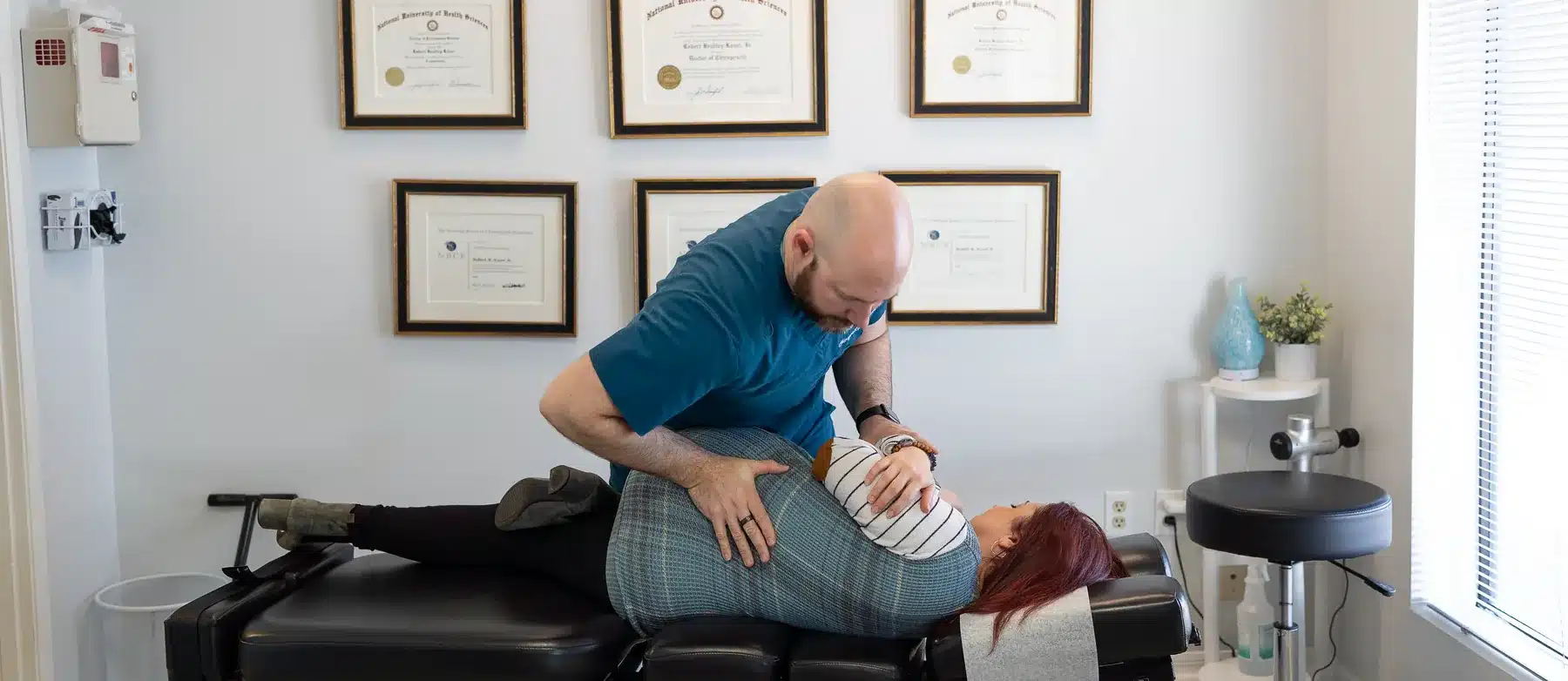
It is 11pm, the report came back this afternoon, and the word on it is arthritis. Maybe with company: joint space narrowing, osteophytes, subchondral sclerosis, degenerative changes. It was written for other doctors, not for you, so at 11pm the internet is reading it to you instead, and the internet is not good at this. This page is the translator. Dr. Bobby Koser, DC, chiropractic physician, examines the joint, the joints above and below it, and how you load it, and his post-doctoral training in spinal MRI interpretation and clinical correlation covers the spine side of a report. Bring yours.
Arthritis Overview
What the words on your report mean
Arthritis is not one condition. It is a word that covers a large family, and the member of that family that report language like joint space narrowing, osteophytes and subchondral sclerosis is describing is osteoarthritis. It shows up in the knees, the hips, the hands, the shoulders, and the spine, and much of the vocabulary a radiologist uses to describe it carries across all of them. Learn the words once and your next report reads faster.
Take a joint apart in your head first. Two bone ends meet. Each end is capped with cartilage, a slick, rubbery coating that lets the surfaces glide across each other. A capsule wraps the whole thing and holds a small amount of fluid inside it. Ligaments hold the two bones in relationship, and muscles and tendons move them and steady them. Osteoarthritis is the name for a set of changes in that structure: the cartilage cap thins or develops defects, the space between the bones on an X-ray gets smaller, the bone at the rim of the joint builds extra ridges, and the bone just under the joint surface changes in ways a scan can see. Your report may describe one of those, or all of them, in one joint or in several.
Osteoarthritis
The form of arthritis that most of the report language on this page is describing. It is a condition of the whole joint rather than of one tissue: cartilage, the bone beneath it, the capsule, the fluid inside it, and the muscles that control it are all part of the picture. A report usually describes several of those features rather than one, and a clinician puts them together with you.
Also written as: OA, osteoarthrosis, degenerative joint disease or DJD, degenerative arthritis, degenerative changes, arthrosis, arthropathy (which means joint condition). Reports name the joint in front of it: knee OA, hip OA, glenohumeral OA at the shoulder, first carpometacarpal OA at the base of the thumb.
Joint space narrowing
Cartilage does not show up on an ordinary X-ray. Bone does. So a radiologist judges the cartilage indirectly, by measuring the gap between the two bone ends, and a smaller gap gets written up as joint space narrowing. Reports may describe how much of it there is in words rather than numbers, and they usually locate it too: medial (the inner side of the knee), lateral (the outer side), or superior (the top of the hip socket). It describes a distance on a picture taken at one moment. What that distance means for you is worked out by examining the joint, because a narrow gap can sit under a joint that works well and a wider one can sit under a joint that hurts.
Also written as: loss of joint space, reduced joint space, narrowing of the medial compartment, cartilage space loss.
Osteophytes, or bone spurs
An osteophyte is a ridge of extra bone that forms along the edge of a joint. Reports call the same thing a bone spur, spurring, marginal osteophyte, or osteophytosis. Where one sits is what the examination looks at: a ridge at the edge of a knee and a ridge crowding the opening a nerve root travels through in the spine are two different conversations, and the spine version has its own page here.
Also written as: bone spur, spurring, osteophyte formation, marginal osteophytes, hypertrophic changes, productive changes.
Subchondral sclerosis, subchondral cysts, bone marrow lesion
Subchondral means under the cartilage, so all three describe the layer of bone directly beneath the joint surface. Sclerosis means that bone reads as denser and brighter on an X-ray. A subchondral cyst is a fluid-filled pocket in that layer. A bone marrow lesion, a term that appears on MRI reports rather than X-rays, is a change in the bone just under the joint surface that shows up bright on the scan. These sit next to each other on reports because they are all describing the same neighborhood.
Also written as: subchondral sclerosis, subarticular sclerosis, geode (an older word for a subchondral cyst), BML, subchondral bone marrow edema pattern.
Cartilage defect, chondral thinning, chondromalacia
Chondral means cartilage. A chondral or cartilage defect is an area where the cartilage cap is thinned or interrupted, and MRI reports often grade how deep it goes, from a surface change to a defect that reaches the bone underneath. Chondromalacia is an older word for softened cartilage, still used most often about the back of the kneecap. Full-thickness means the defect goes all the way through to bone at that spot. How large that spot is, where it sits, and what the joint does when it is loaded are what the examination adds to the word.
Also written as: chondral defect, chondral fissuring, cartilage loss, chondromalacia patellae, grade 2 or grade 3 chondral change, cartilage thinning.
The grade on your X-ray
Knees and hips often come back with a number attached. The Kellgren-Lawrence scale runs 0 to 4 and counts the changes a radiologist can see on the film: joint space narrowing, osteophytes, and changes in the bone near the joint surface. Other scales exist and some reports use words instead of numbers. A grade summarizes a picture. It is not a score for how much you hurt, it is not a prediction, and two people carrying the same grade can be living very different days. What it gives the examination is one more piece of information to line up against what your joint actually does under load.
Also written as: Kellgren-Lawrence grade, KL grade, KL 2, grade 2 osteoarthritis, radiographic grade, Tonnis grade at the hip.
The words that show up on hands
Hands get their own vocabulary because the swellings are visible. Heberden nodes are firm enlargements at the joints closest to the fingertips. Bouchard nodes are the same thing at the middle knuckles. Reports may also name the base of the thumb, written as first carpometacarpal or first CMC osteoarthritis, the joint that makes jar lids, keys and car doors a daily negotiation. Erosive osteoarthritis is a less common pattern in the fingers, one where the report describes erosions, meaning areas where bone at the joint surface has been lost, and that word on a hand report is one of the reasons a visit goes to a physician for blood work rather than straight into a care plan.
Also written as: DIP and PIP joint osteoarthritis, nodal osteoarthritis, thumb base arthritis, basal joint arthritis, trapeziometacarpal osteoarthritis.
When the report is about your spine
The spine uses a parallel vocabulary for the same family of changes, because the small joints at the back of each spinal segment are joints like any other: facet arthropathy, facet hypertrophy, spondylosis, degenerative joint disease of the lumbar or cervical spine. Those words, and what they do and do not tell you, are on our spinal arthritis and facet joint pain page.
Also written as: facet arthropathy, facet arthrosis, facet hypertrophy, spondylosis, degenerative joint disease of the lumbar or cervical spine, facet joint osteoarthritis.
A finding is not a diagnosis
Here is something worth knowing before you read your report again, and it has been measured in more than one joint.
Start with the hip. One study compared hip pain against hip X-rays in two separate groups of people, 946 in the first group and 4,366 in the second. Most hips that hurt on most days did not show osteoarthritis on the X-ray: 15.6% did in the first group and 9.1% in the second. It ran the other way too. Of the hips that did show osteoarthritis on the X-ray, 20.7% in the first group and 23.8% in the second were frequently painful. Most of the older participants whose pain pattern or painful hip rotation raised a high suspicion of hip osteoarthritis did not have it on the X-ray, which suggested to the authors that in many cases hip osteoarthritis might be missed if the diagnosis rested on the X-ray alone.1 Both halves matter. A finding on your report is not automatically the cause of your pain, and your pain is not less real because the X-ray looks unremarkable. The two groups were analyzed separately, the study asked participants whether their hip hurt on most days rather than counting hips in the days after a crash or a fall, the films were X-rays rather than MRI scans, and the study compared pictures and reported pain at a single point in time, so nothing in it speaks to what came first or what happens next. More on the hip specifically is on our hip pain page.
The knee has been counted the same way. In a review of 63 studies covering 5,397 knees from 4,751 adults who had no knee symptoms and no knee injury, MRI scans showed cartilage defects in about 24%, bone spurs, which reports call osteophytes, in about 25%, bone marrow lesions, a change in the bone just under the joint surface that shows up bright on the scan, in about 18%, and tears in the meniscus, the C-shaped cushion between the thigh bone and the shin bone, in about 10%.2 In that review, again in adults with no knee symptoms and no knee injury, the summary estimates across those findings ran from 4% to 14% in adults under 40 and from 19% to 43% in adults 40 and older, depending on which finding, and the reviewers noted that osteophytes became more common with age while bone marrow lesions did not. That cuts in both directions, and both matter. A finding on your report is not automatically the cause of your pain, and your pain is not less real because the same words appear on other people’s reports. In that review the estimates also moved with how the MRI scans were done, with physical activity, with whether osteoarthritis was already visible on X-ray, and with the risk of bias in the studies pooled, so they are pooled estimates rather than fixed rates. And every knee in it was an uninjured knee, which is why nothing in those figures describes a knee after a crash, a fall, or a sports injury. Our knee pain page goes further into the knee.
Then there is the sound, which is the question people actually ask first. In a review of 103 studies covering 42,816 knees, knee crepitus, the crackling or grinding noise a knee can make when it moves, was reported in about 41% of the general population, a figure drawn from 7,609 knees, in about 36% of pain-free knees, a figure drawn from 852 knees with a 95% confidence interval of 23% to 50%, and in about 81% of knees with osteoarthritis, a figure drawn from 18,821 knees. The same review counted crepitus in about 35% of knees with a ligament injury, a figure drawn from 2,740 knees, and in about 61% of knees with cartilage pathology, meaning a problem with the cartilage itself, a figure drawn from 1,445 knees, so a noise that started after an injury is its own question. In that review crepitus was also associated with more than threefold higher odds of osteoarthritis on X-ray, an odds ratio of 3.79 with a 95% confidence interval of 1.99 to 7.24, a figure drawn from 1,725 knees, a comparison between groups and its range of statistical uncertainty rather than a number about any one person.3 The reviewers graded the certainty of these estimates as low to very low and said they should be read with caution. So the sound alone does not settle the question in either direction, which is what the examination is for.
Put all three together and the same shape appears. A report can be accurate and still not be the answer to your question. Whether a finding explains your symptoms depends on where it sits, what it sits next to, whether it matches what you feel and where you feel it, and what the joint does when it is loaded and moved in front of somebody. The report is the starting point. The examination connects it to you.
1. Kim C, et al. Association of hip pain with radiographic evidence of hip osteoarthritis: diagnostic test study. BMJ. 2015;351:h5983. PubMed
2. Culvenor AG, et al. Prevalence of knee osteoarthritis features on magnetic resonance imaging in asymptomatic uninjured adults: a systematic review and meta-analysis. Br J Sports Med. 2019;53(20):1268-1278. PubMed
3. Couch JL, et al. Noisy knees. Knee crepitus prevalence and association with structural pathology: a systematic review and meta-analysis. Br J Sports Med. 2025;59(2):126-132. PubMed
When it is not osteoarthritis
The word arthritis covers a second family that behaves differently and is managed along a different path. Rheumatoid arthritis, psoriatic arthritis, gout, and reactive arthritis are inflammatory conditions, meaning the pain and swelling are driven by an inflammatory process rather than by changes in the joint structure. They are diagnosed with a physician’s workup, usually blood work and sometimes fluid drawn from the joint, and they are treated with medication a chiropractic physician does not prescribe. Getting a person into that room promptly is the point, because that is where the treatment for those conditions starts.
So the examination here asks a set of questions whose whole purpose is to find out whether you belong on the osteoarthritis path at all. None of the patterns below is a diagnosis. Each is a reason to get you in front of your primary care physician or a rheumatologist, the physician who specializes in arthritis and autoimmune conditions.
- Stiffness in the morning that runs past an hour. How long you are stiff before the joint loosens up is one of the more useful things you can time for yourself. Stiffness measured in hours turns the conversation toward a physician.
- A joint that is hot, visibly swollen, or red. Warmth you can feel with the back of your hand is not something to work around at an appointment. A hot joint with fever is an emergency department visit now.
- The same small joints on both sides at once. Knuckles, wrists, the balls of the feet, matching left and right, is a pattern that belongs in front of a physician.
- Symptoms that are not confined to the joint. Fever, unexplained weight loss, night sweats, a rash, mouth ulcers, eye redness or pain, or fatigue that is out of proportion to the day you had.
- A single joint that goes from fine to painful over hours rather than over weeks, often the base of the big toe, sometimes a knee or an ankle, sometimes with the skin tight and shiny over it. That tempo is one a physician evaluates, with testing that happens there rather than here.
- Psoriasis, inflammatory bowel disease, or a family history of an inflammatory arthritis, and joint pain on top of it.
- Joint pain that started in the weeks after a gut or urinary infection.
- Back pain that started before age 40, came on gradually, wakes you at night, and eases with movement rather than with rest. That combination is covered on our spinal arthritis page, where the research on the pattern is set out.
People also arrive already carrying an inflammatory diagnosis and a rheumatologist, and with the mechanical parts of their day still difficult: a stiff mid back, a shoulder that will not reach the shelf, muscles that have been guarding a joint for months. That work happens alongside their physician’s care rather than instead of it, and it starts with an examination.
Symptoms, and the warning signs we screen for
Osteoarthritis is usually noticed through a joint’s relationship with load. Pain that comes up with use and settles with rest. Stiffness first thing in the morning, or after a long meeting or a long drive, that eases within a few minutes of moving. A joint that grinds, crackles or catches. Swelling that appears after a busy day. Less range than the other side has, so the shoulder stops short of the top shelf, the hip will not let you cross your legs to put a sock on, the knee objects to stairs and to getting out of a low car seat, and the thumb loses its argument with a jar lid.
Which joint is involved changes what the examination checks, but not the order of operations. A painful knee is examined with the hip and the foot, because the knee sits between them and takes what they hand it. A painful hip is examined with the low back, because the two trade symptoms constantly. A painful shoulder is examined with the neck and the shoulder blade. A hand is examined with the wrist, the elbow and the neck. If you have more than one joint involved, that changes the questions the visit asks, including the ones in the section above.
Common, and examined here
- Knee, hip, hand, shoulder or back pain that is worse with use and eases with rest
- Stiffness in the morning or after sitting that loosens as you move
- A joint that grinds, crackles or catches
- Swelling in a joint that comes up after activity
- Trouble with stairs, a low car seat, a jar lid, or reaching overhead
- An X-ray or MRI report saying osteoarthritis, degenerative changes, or bone spurs that nobody has explained to you
Emergency, not an appointment
- A hot, swollen joint with fever
- A joint that will not take your weight after an injury
- New swelling or pain in the calf, more urgent still with chest pain or shortness of breath
Go to the emergency department now. A hot joint with fever can mean infection inside the joint, which is treated as an emergency. New swelling or pain in the calf can mean a blood clot in the leg, and chest pain or shortness of breath along with it makes it more urgent still. This is part of what the examination here screens for.
How we approach arthritis
The examination comes first: your history, the joint taken through its range and compared with the other side, the muscles and tendons that cross it tested directly, strength and load tolerance measured, the joints above and below it examined in the same visit, and any report you already have read with you rather than at you. That visit is the Pain Source Exam. The question it answers is the one you came in with: does what the picture shows explain the person in the room, and if it does, what part of your day is it actually costing you?
Bring films and reports if you have them. If you do not, the examination decides whether imaging is worth ordering. Imaging is ordered when the examination raises a question a picture can answer: an injury that could have broken something, a joint that will not take weight, a joint that locks or gives way, findings that do not line up with the story, a pattern that could be inflammatory or infectious and needs a physician’s workup around it, or a case heading toward a surgical opinion. Where the examination already answers the question, the visit spends its time on the examination.
When conservative care fits, it is built from what the examination found rather than from what the report said. That means a chiropractic adjustment where the examination finds restricted motion, in the joint itself or in the joints feeding it, with the technique chosen for the joint in front of us. Hands-on muscle work for the tissue that has been guarding the area. Dry needling for trigger points, meaning tight, painful knots in a muscle, or myofascial pain, meaning pain from the muscle and its covering tissue, in the muscles around the joint. And a movement plan, meaning specific loading prescribed at a dose you can complete and reviewed at the next visit, because a joint that is stiff and a joint that is irritable want different doses, and yours is found by prescribing one, doing it, and checking it at the next visit. Care is monitored, and if you are not progressing, the plan changes.
Some joints are examined here and then sent elsewhere.
- A pattern that behaves like inflammatory arthritis rather than osteoarthritis: your primary care physician or a rheumatologist, with what the examination found sent along.
- A hot, swollen joint with fever: emergency department, not an appointment.
- A joint that locks, catches hard, gives way, or will not take weight after an injury: orthopedic opinion and imaging first.
- A joint where a joint replacement is on the table: an orthopedic surgeon, and that decision belongs with them.
- A child or adolescent with a limp and knee, thigh or hip pain: pediatric orthopedics the same day, because a hip problem at that age is often felt at the knee.
- A joint that is not progressing under a monitored plan: re-examined here, and referred for a medical or orthopedic opinion if the examination points there.
When we refer you, your records go with you. Hurt in a car accident? Florida law gives you 14 days from the crash to begin care and keep your no-fault medical benefits. If that window is open, start at our auto accident page and we will get you examined.
What the research shows
Two pieces of research come up when people are told what to do about arthritis: one on movement and body weight, one on hands-on care.
In an 18-month trial, 454 adults aged 55 and older whom the trial’s entry criteria classified as overweight or obese, with a body mass index between 27 and 41, and who had knee pain along with osteoarthritis on X-ray, were assigned to an intensive diet plus exercise, to the diet alone, or to exercise alone.4 In that trial the diet-plus-exercise group lost about 10.6 kg on average, roughly 23 pounds, which was about 11% of body weight, and at 18 months reported less pain and better function than the exercise-only group. Put the other way, in that trial the diet-only group’s pain score, 4.8 on a scale that runs 0 to 20, was about the same as the exercise-only group’s 4.7, and the trial reported no advantage for the diet alone over exercise alone on that score, so what the trial separated out was the combination of the two. Pain, function and quality of life were secondary outcomes there, meaning results the trial collected alongside the ones it was built to measure, and the trial’s main measurements were laboratory and mechanical ones rather than how people felt. Everyone enrolled was 55 or older with knee pain and X-ray changes together, at a single university center running a center-based diet and exercise program, with the exercise groups able to move to a home-based program. It is a knee trial, so it says nothing about a hip, a shoulder or a hand. What happens with it here is an examination that finds what your joint tolerates and builds the movement side of a plan from there, with questions about diet and body weight belonging to you and your primary care physician.
Hands-on care has been reviewed too, and the review is small and old: a systematic review published in 2011 searched for randomized trials of manual therapy in hip or knee osteoarthritis, meaning trials where chance decided who got which kind of care, and found four, 280 people in all.5 Three of those four studied the knee and one studied the hip. Three of the four were judged by the reviewers to be at high risk of bias, the studies differed enough that the reviewers could not pool them, and the reviewers wrote that this evidence on manual therapy for pain and function in knee or hip osteoarthritis could be considered inconclusive, because there were so few trials and so few patients. The reviewers also reported that the one hip trial favored manual therapy over exercise in both the short and the long term, on evidence they labelled silver level, which is their grading term for how strong the evidence is rather than a measure of how much better it was. All four reported short-term effects and one measured long-term effects. That review does not cover work published since it was written. That is why hands-on work here sits inside a plan that gets re-examined at the next visit.
In both of those, what you are reading is what a study found in the people it enrolled, not a prediction for your joint. Finding out where you actually stand is what the examination is for.
4. Messier SP, et al. Effects of intensive diet and exercise on knee joint loads, inflammation, and clinical outcomes among overweight and obese adults with knee osteoarthritis: the IDEA randomized clinical trial. JAMA. 2013;310(12):1263-1273. PubMed
5. French HP, et al. Manual therapy for osteoarthritis of the hip or knee. A systematic review. Man Ther. 2011;16(2):109-117. PubMed
Common questions about arthritis
My X-ray says osteoarthritis. Is that why I hurt?
Not automatically, and it matters in both directions. That question has been measured directly in the hip. One study compared hip pain against hip X-rays in two separate groups of people, 946 in the first group and 4,366 in the second. Most hips that hurt on most days did not show osteoarthritis on the X-ray, 15.6% did in the first group and 9.1% in the second, and of the hips that did show it, 20.7% in the first group and 23.8% in the second were frequently painful. Most of the older participants whose pain pattern or painful hip rotation raised a high suspicion of hip osteoarthritis did not have it on the X-ray, which suggested to the authors that in many cases hip osteoarthritis might be missed if the diagnosis rested on the X-ray alone. A finding on your report is not automatically the cause of your pain, and your pain is not less real because the X-ray looks unremarkable. The study asked participants whether their hip hurt on most days rather than counting hips in the days after a crash or a fall, the films were X-rays rather than MRI scans, and the pictures and the pain were compared at a single point in time. Whether the finding explains you is what an examination works out.
What is the difference between osteoarthritis and rheumatoid arthritis?
They share a word and very little else. Osteoarthritis describes changes in the structure of a joint: thinner cartilage, a smaller space between the bones on an X-ray, extra bone at the rim, and changes in the bone just under the joint surface. Rheumatoid arthritis is an inflammatory condition, meaning an inflammatory process drives the pain and swelling, and it tends to involve the same small joints on both sides with stiffness that runs past an hour in the morning. Rheumatoid arthritis is diagnosed with a physician’s workup, usually blood work, and treated with medication a chiropractic physician does not prescribe, which is why that pattern gets referred rather than managed here. Psoriatic arthritis, gout and reactive arthritis sit in the same inflammatory family and follow the same route.
My knee makes noise. Does that mean I have arthritis?
The noise alone does not settle it either way. In a review of 103 studies covering 42,816 knees, knee crepitus, the crackling or grinding noise a knee can make when it moves, was reported in about 41% of the general population, a figure drawn from 7,609 knees, in about 36% of pain-free knees, a figure drawn from 852 knees with a 95% confidence interval of 23% to 50%, and in about 81% of knees with osteoarthritis, a figure drawn from 18,821 knees. The same review counted crepitus in about 35% of knees with a ligament injury, a figure drawn from 2,740 knees, and in about 61% of knees with cartilage pathology, meaning a problem with the cartilage itself, a figure drawn from 1,445 knees, so a noise that started after an injury is its own question. In that review crepitus was also associated with more than threefold higher odds of osteoarthritis on X-ray, an odds ratio of 3.79 with a 95% confidence interval of 1.99 to 7.24, a figure drawn from 1,725 knees, a comparison between groups and its range of statistical uncertainty rather than a number about any one person. The reviewers graded the certainty of those estimates as low to very low and said to read them with caution. So a noisy knee is a question the examination answers, by looking at what else the knee does, not something a page can answer for you.
Will losing weight fix my knees?
One trial is usually what people are quoting, and what it credited was diet and exercise together. In an 18-month trial, 454 adults aged 55 and older whom the trial’s entry criteria classified as overweight or obese, with a body mass index between 27 and 41, and with knee pain along with osteoarthritis on X-ray, were assigned to an intensive diet plus exercise, to the diet alone, or to exercise alone. In that trial the diet-plus-exercise group lost about 10.6 kg on average, roughly 23 pounds, which was about 11% of body weight, and at 18 months reported less pain and better function than the exercise-only group. Put the other way, the diet-only group’s pain score, 4.8 on a scale that runs 0 to 20, was about the same as the exercise-only group’s 4.7, and the trial reported no advantage for the diet alone over exercise alone on that score, so what the trial separated out was the combination of the two. Pain and function were secondary outcomes there, meaning results the trial collected alongside the ones it was built to measure, and everyone in it was 55 or older with knee pain and X-ray changes together, at one university center running an 18-month program. That is what a trial found in the people it enrolled. Diet is a conversation for you and your primary care physician; the movement side is part of what an examination here sets.
Should I keep moving, or rest it?
That is a dose question rather than a yes-or-no question, and it is one of the things the examination is for. A joint that is stiff and a joint that is irritable respond to different amounts of loading, and the amount that suits you is found by prescribing a specific dose, doing it, and checking it at the next visit rather than by guessing at the start. What that means in practice is that your plan is written to be reviewed and changed, and that you leave with something to do between visits.
What does chiropractic care actually do for arthritis?
The examination looks at the joint, the joints above and below it, the muscles crossing it, and how you load it during your day, and it decides whether a mechanical problem is part of what you are feeling. Where it is, care addresses that part: restricted motion in the joint or the joints feeding it, muscle that has been guarding the area, and a loading program you can complete. On the research, a systematic review published in 2011 found four randomized trials of manual therapy in hip or knee osteoarthritis, meaning trials where chance decided who got which kind of care, 280 people in all, and the reviewers wrote that this evidence could be considered inconclusive because there were so few trials and so few patients. Those same reviewers also reported that the one hip trial in that review favored manual therapy over exercise in both the short and the long term, on evidence they labelled silver level, which is their grading term for how strong the evidence is rather than a measure of how much better it was. That review does not cover work published since it was written. Care here is monitored for that reason, and if you are not progressing, the plan changes.
Do I need an X-ray or MRI before I come in?
No. If you already have films or a report, bring them, because reading them with you is part of the visit. If you do not have them, the examination decides whether imaging is worth ordering. It gets ordered when the examination raises a question a picture can answer, such as an injury that could have broken something, a joint that will not take weight, a joint that locks or gives way, findings that do not line up with your story, or a case heading toward a surgical opinion.
When is joint pain an emergency?
A hot, swollen joint with fever, which can mean infection inside the joint. A joint that will not take your weight after an injury. New swelling or pain in the calf, which can mean a blood clot in the leg, and more urgent still if chest pain or shortness of breath comes with it. Those are emergency department visits now, not appointments.
 Top 3 Finalist: Best Chiropractor
Creative Loafing Best of the Bay 2025 · Readers Pick
Quoted in National Press
See the coverage →
Top 3 Finalist: Best Chiropractor
Creative Loafing Best of the Bay 2025 · Readers Pick
Quoted in National Press
See the coverage →
This page is general health information and is not medical advice for any individual. The studies summarized describe groups, not any one person’s outcome, and whether any finding on your imaging explains your symptoms can only be determined by examination. Talk to Dr. Bobby Koser, DC, about your own situation, and seek emergency care immediately for the warning signs listed above.
Bring the report, and the joint it is about.
A complete examination with Dr. Bobby Koser, DC, your films and reports reviewed with you, and your findings explained the same day.